
Presentation
Presented with sever pain and deformity of the upper left thigh. Chronic history of left hip pain with progressive difficulty walking. No prior history of trauma.
Patient Data
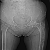
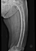

Diffuse cortical and coarse trabecular thickening with bowing of left femur.
Multifocal osteolytic defect along the cortex.
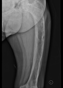
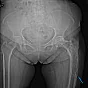
Transverse insufficiency fracture is known as a "banana fracture" at the lateral cortex of the femur.

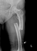
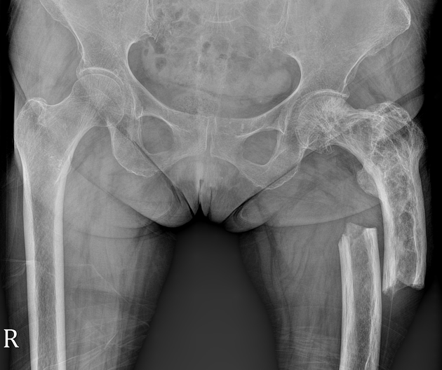
Displaced complete transverse pathological fracture at the same site of previously mentioned lateral cortex insufficiency fracture.
Diffuse cortical and coarse trabecular thickening with bowing of left femur.
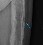

Annotated and magnified images for transverse insufficiency banana fracture (arrow) at the lateral cortex of the left femur.
Case Discussion
Paget disease of bone is a benign disease characterized by exaggerated remodeling of the bone matrix after osteoclast-mediated bone destruction. Its etiology is still unknown.
The main clinical presentations of the disease are related to bone pain and deformities.
Radiological diagnosis is the main detection tool, though many monostotic Paget disease cases may remain undiagnosed.


 Unable to process the form. Check for errors and try again.