
Presentation
A female presented with a history of generalized bone pain. Serum calcium and parathyroid hormone are elevated(Ca: 11.5 mg/dl and PTH: 118 pg/mL).
Patient Data
Age: 50 years
Gender: Female
From the case:
Parathyroid adenoma

Download
Info
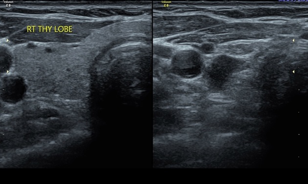
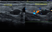
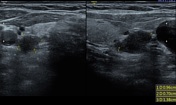
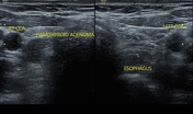
There is a well-defined, homogeneous lesion posterior-inferior to the right lobe of the thyroid gland.
It is hypoechoic in comparison to the thyroid gland with no evidence of cystic changes or internal calcifications
Few flow signals are present in the lesion.
Case Discussion
She was referred for a neck ultrasound to rule out/rule in a parathyroid adenoma. Regarding the clinical, laboratory, and imaging findings, the parathyroid adenoma is the final diagnosis. Surgical excision was done and histopathology confirmed the lesion being a parathyroid adenoma.


 Unable to process the form. Check for errors and try again.