
Presentation
Prior FOOSH and resultant intraarticular fracture of the distal radius sustained and operated elsewhere. Ongoing pain and limited movement of the radiocarpal joint after 3 months.
Patient Data
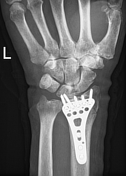


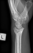
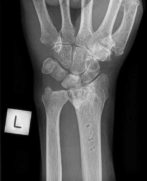
Status post volar locking plate fixation. The fracture lines are still visible. The two ulnarmost screws seem to protrude into the radiocarpal joint. Subtle periarticular patchy osteopenia suggestive of complex regional sympathetic dystrophy.
Stat CT was recommended for further assessment.


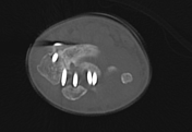
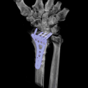
Partial nonunion of the fracture and protrusion of the two ulnarmost screws into the joint, which are abutting the lunate. Patchy regional osteopenia indicating reflex sympathetic dystrophy are clearly depicted.
The patient was reoperated shortly afterwards.

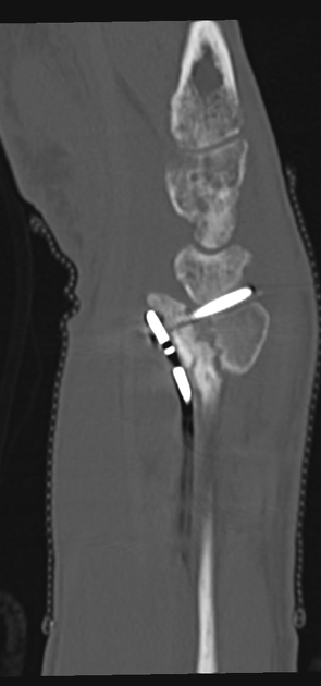
Posttraumatic progressive degenerative changes are visible in the radiocarpal joint.
Case Discussion
Careful assessment of screw position and comparison with prior radiographs is crucial while assessing any sort of screw fixation device. Inherently poor position or postoperative migration of screws into the joint space can be a cause of severe pain, and jeopardizes the long term outcome. The correct position of the radial metaphyseal shaft screws is also critical and these should not extend significantly beyond the dorsal cortex, where they could abut extensor tendons.


 Unable to process the form. Check for errors and try again.