
Presentation
Vaginal mass on gynecological exam.
Patient Data
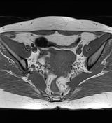

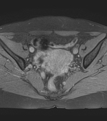
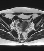
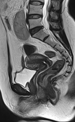
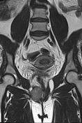
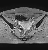
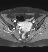
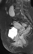
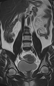
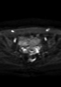
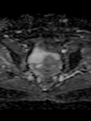

The MRI sequences demonstrate a well-defined ovoid right vaginal soft tissue mass (40 x 37 x 35 mm) centered on the Bartholin gland, displacing the puborectalis laterally and anorectal region medially. It elicits a slight high signal on T1 and TI fat sat, intermediate to low signal on T2 with restricted diffusion and a homogeneous enhancement postcontrast sequences.
Note ectopic malrotated left kidney.
Conclusion (pathological report):
Morphological appearance and immunochemistry study of a vaginal pleomorphic leiomyosarcoma.
Case Discussion
MRI features of a vaginal tumor.
The patient underwent a complete resection of the tumor with histopathological exam and immunochemistry study which were in favor of a vaginal pleomorphic leiomyosarcoma.


 Unable to process the form. Check for errors and try again.