
Presentation
None provided.
Patient Data
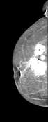

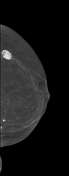
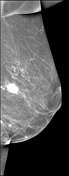

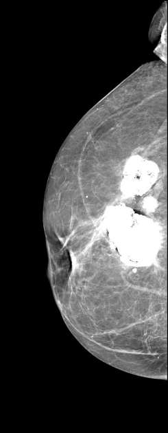
Cluster of coarse calcifications in the right breast at 11-12 o'clock.
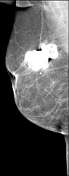
Single coarse calcification in the left breast at 3 o'clock.
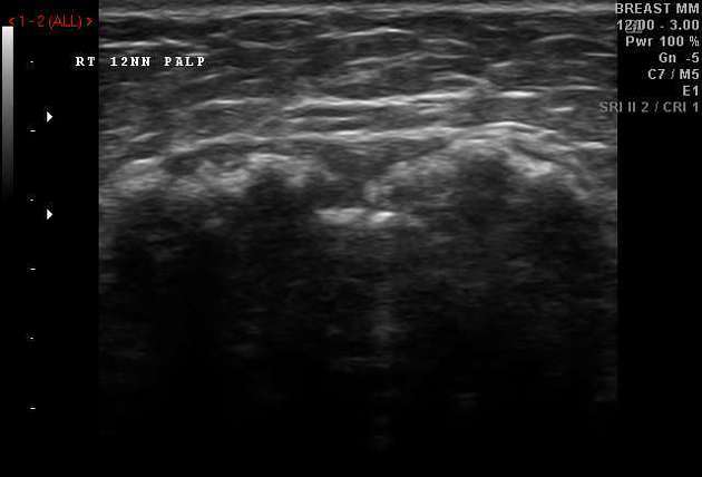
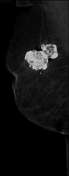
Coarse calcifications corresponding to the positions of those on the mammograms.
Case Discussion
The calcified masses were identified as foci of fat necrosis and remained unchanged for years. Nonetheless, Tru-cut biopsies were obtained 5 years after the above mammography and ultrasound studies:
Right breast, 12 o'clock - calcified mass, most probably fat necrosis.
Macroscopic description:
Yellow crumbs of tissue, 1.0-1.1 cm in diameter. Marked with hematoxylin.
Microscopic description:
Cores of fibro-fatty tissue with signs of fat necrosis, calcifications.
Left breast, 3 o'clock - calcified mass, most probably fat necrosis.
Macroscopic description:
Yellow crumbs of tissue, 0.2-0.3 cm in diameter. Marked with hematoxylin.
Microscopic description:
Cores of fibro-fatty tissue with signs of fat necrosis, calcifications.


 Unable to process the form. Check for errors and try again.