
Presentation
Workup for right-sided hydronephrosis that is reported in recent ultrasonography.
Patient Data
Age: 10 years
Gender: Female
From the case:
Retrocaval ureter - type I


Download
Info
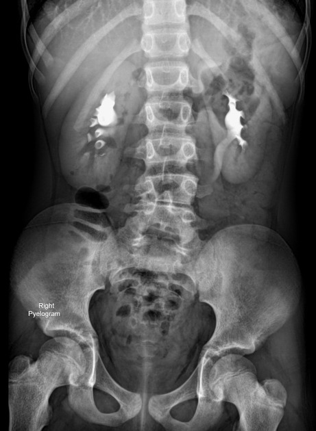
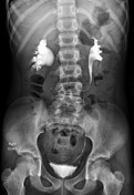
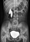
The right kidney shows hydronephrosis, and there is right proximal ureteric dilatation with medial deviation and an abrupt change in mid-ureter caliber (fishhook-shaped or S-shaped deformity) without detectable radio-opaque shadows along the course of the urinary tract.
Case Discussion
Features are compatible with the retrocaval ureter.
Retrocaval ureter, also known as circumcaval ureter, is a rare congenital anomaly in which the right ureter passes behind the inferior vena cava, then emerges between the IVC and aorta and passes anterior to the IVC.


 Unable to process the form. Check for errors and try again.