
Presentation
Psa 23.4. DRE - T2 right lobe with bilateral firmness throughout. Are there any other areas to target at biopsy?
Patient Data


Prostate volume 37mls. PSA density is 0.63ng/ml.
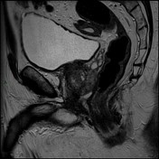
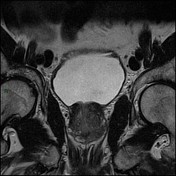
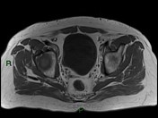
1.6 cm irregular low T2 signal abnormality centered on the right peripheral zone at the gland apex. This extends into the transitional zone and across the midline. Irregularity of the prostatic capsule. Corresponding b1400 changes ADC restriction. (PIRADS 5).
2.2 cm low signal lesion in the posterolateral aspect of the left peripheral zone at the level of the mid gland and extending towards the apex. This breaches the capsule and abuts the puborectalis muscle, however without overt invasion (PIRADS 5).
Normal seminal vesicles.
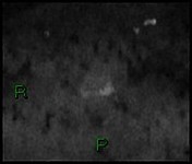
Suspicious 7 mm left obturator node. No sinister bone lesion.
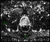
Matrix of the Lt PIRADS 5...
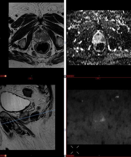
Matrix of the Lt PIRADS 5 lesion
A matrix illustrating the left PIRADS 5 lesion.
Focal lesions are typically labeled/annotated/marked up for the clinician undertaking the biopsy.
Most centers would undertake a biopsy using transrectal ultrasound guidance.
Case Discussion
Appearances in keeping with bilateral prostate malignancy.
Radiological stage T3a, N1, MX.
Transrectal ultrasound biopsy (TRUS) was performed.
Histology: prostate adenocarcinoma
Right lobe: Gleason 9 (4+5). 70% of tissue. Perineural invasion.
Left lobe: Gleason 9 (4+5). 90% of tissue. Perineural invasion.


 Unable to process the form. Check for errors and try again.