
Presentation
Abdominal pain and mass feeling on physical exam.
Patient Data
Age: 50 years
Gender: Female
From the case:
Giant hepatic hemangioma


Download
Info

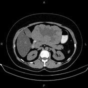
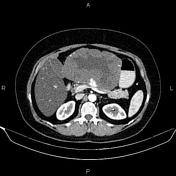
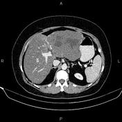
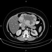
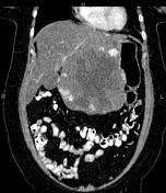
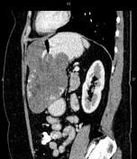
The hepatic attenuation value is less than the spleen's, suggesting fatty liver.
A 140×95 mm hetero-dense lesion at the left liver lobe shows early peripheral nodular enhancement with centripetal filing and delayed blood pools.
Case Discussion
Features are typical for giant hepatic hemangioma. It is essential to differentiate giant hemangiomas from hepatic malignancies such as hepatocellular carcinoma and hepatic metastases.


 Unable to process the form. Check for errors and try again.