
Presentation
Known case of uterine mass. Acute pelvic pain for one week.
Patient Data
Age: 25 years
Gender: Female
From the case:
Uterine fibroid with internal degeneration


Download
Info
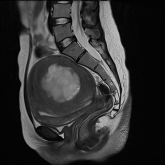
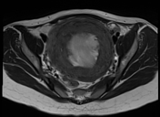
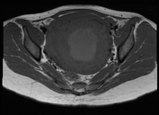
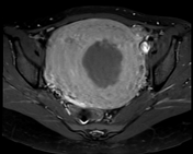
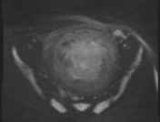
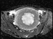
A large peripherally solid enhancing mass lesion with central non-enhancing (T1 intermediate and T2 hyperintense signal) in the posterior wall of the uterus measuring about 10 x 10 x 11 cm.
The endometrial cavity is compressed inferiorly by the lesion without evidence of distention.
No surrounding aggressive features.
No extension beyond the uterine contour.
No enlarged pelvic lymph node. No significant pelvic ascites.
Case Discussion
The features are suggestive of uterine mural large fibroid with internal degeneration.


 Unable to process the form. Check for errors and try again.