
Presentation
G3P2. at 20 weeks gestation. Routine ultrasound exam.
Patient Data
Age: 35 years
Gender: Female

Download
Info
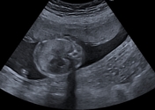
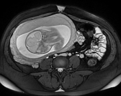
The ultrasound exam:
- expanded right upper lobe with markedly increased echogenicity
- compressed with decreased echogenicity of the right lower lobe and probably the middle lobe
- leftward shift of the heart and mediastinal structures
- compressed left lung with reduced volume
No other fetal anomalies were seen.
Download
Info
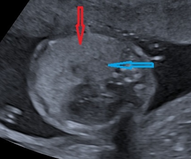
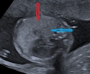
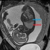
Annotated images:
- red arrow: expanded hyperechogenic right upper lobe
- blue arrow: compressed right lower lobe and probably the middle lobe


Download
Info

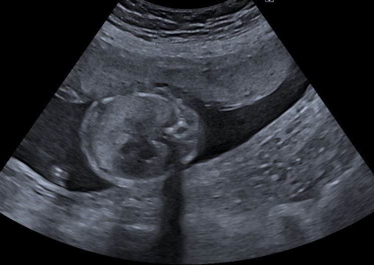
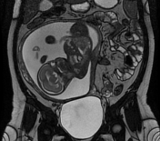
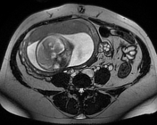
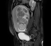
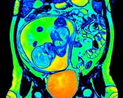
The MRI sequences demonstrate:
- increased volume with hyperintensity of the affected right upper lobe
- compressed with decreased intensity of the right lower lobe and probably the middle lobe
- mass effect with mediastinal shift to the left
- preserved lung architecture
- decreased volume with hypointensity of the normal compressed left lung
No other associated fetal malformations were seen.
Normal amount of amniotic fluid.
Placenta corporeal/fundal anterior 9 cm from the internal os.
Download
Info

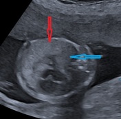
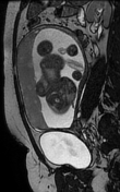
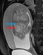
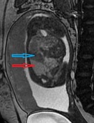
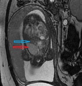
Annotated images:
- red arrow: expanded hyperintense right upper lobe
- blue arrow: compressed hypointense right lower lobe and probably the middle lobe
Case Discussion
Ultrasound and MRI showing:
- expanded right upper lobe with increased echogenicity/intensity
- compressed with decreased echogenicity/intensity of the right lower lobe and probably the middle lobe
- mass effect with a leftward shift of the heart
- compressed with a reduced volume of the normal left lung.
These findings are suggestive of a congenital lobar overinflation.
On imaging, MRI is relatively superior to ultrasound to differentiate between congenital lobar overinflation from pulmonary sequestration and microcystic adenomatoid malformation 1.
Additional contributor: Dr: A. Ounissi, gynecologist, Khenchela, Algeria


 Unable to process the form. Check for errors and try again.