
Presentation
Pelvic pain and mass feeling.
Patient Data
Age: 50 years
Gender: Female
From the case:
Uterine leiomyosarcoma


Download
Info

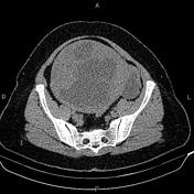
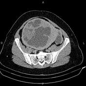
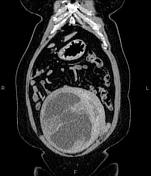
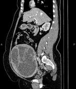
A 195×145×160mm large well defined hetero enhancing solid cystic mass is noted at the right aspect of the uterus body and the fundus, displacing the endometrial cavity to the left side.
Additionally, a 48×34mm cystic lesion without a prominent enhancing solid component is noted on the left side.
The hepatic attenuation value is less than the spleen, suggesting fatty liver disease.
A 20mm gallstone is present.
Case Discussion
The patient underwent a total abdominal hysterectomy, bilateral salpingo oophorectomy, and histopathology evaluation confirmed uterine leiomyosarcoma, a generally poor prognosis malignant uterine tumor that arises from the myometrium.


 Unable to process the form. Check for errors and try again.