
Presentation
Knee pain for 2 weeks. ACL repair performed 2 yrs ago. No acute history of trauma or excessive physical activity.
Patient Data
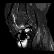

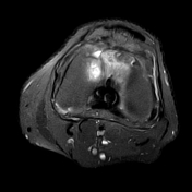
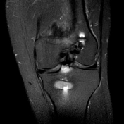
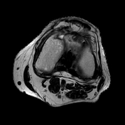
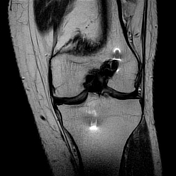
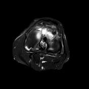
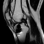
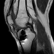
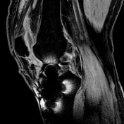

A 2.3 x 3 x 1.4 cm (AP x TR x CC) T2/STIR hyperintense,T1 intermediate well defined soft tissue lesion is seen anterior to the ACL graft - in a case post ACL reconstruction, likely represents cyclops lesion/arthrofibrosis.
Intact ACL graft with mild laxity near its femoral attachment. Clinical evaluation is advised for any signs of laxity.
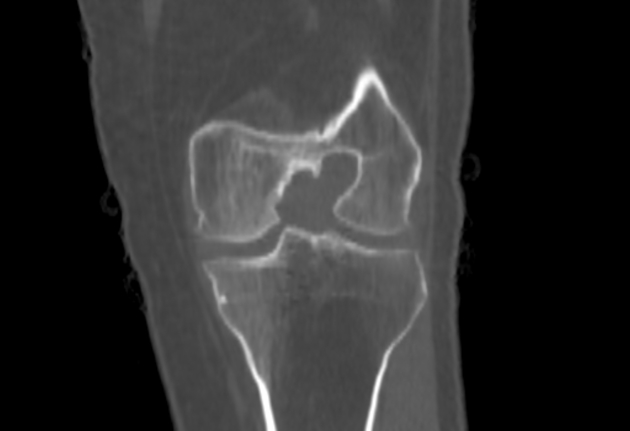
Mild joint effusion with marrow edema in distal femoral shaft.
Rest of the structures are normal.
Case Discussion
A cyclops lesion is localized arthrofibrosis anterior to the ACL graft. It may present with knee pain or reduced range of movement.
The differential diagnosis for a cyclops lesion includes tenosynovial giant cell tumor, synovial chondromatosis and intra-articular loose bodies.
However with the operative history known, the diagnosis is clear and certain.


 Unable to process the form. Check for errors and try again.