
Presentation
Abdominal pain and elevated liver enzymes.
Patient Data
Age: 45 years
Gender: Female
From the case:
Giant hepatic hemangioma


Download
Info
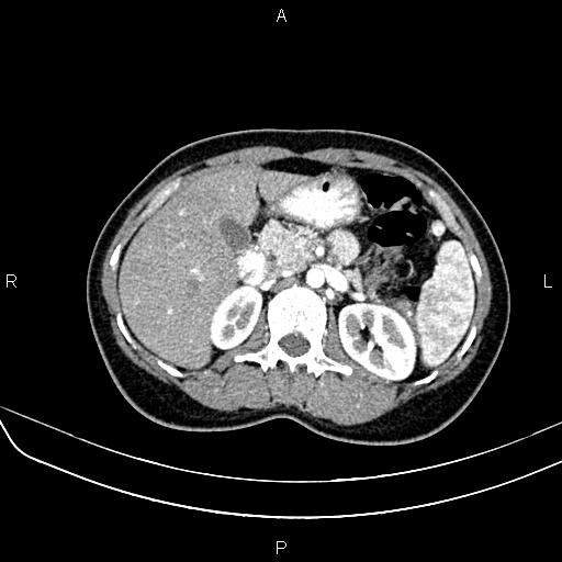
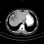
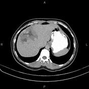
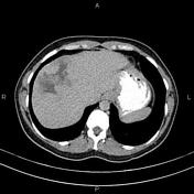
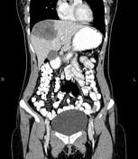
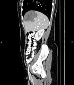
There is an 85 × 75 mm hypoattenuating mass in the liver that shows early peripheral nodular discontinuous enhancement with centripetal filling and delayed blood pools. Large central necrotic components are also evident.
Case Discussion
This case shows a hepatic mass compatible with a giant hepatic hemangioma, also known as giant hepatic venous malformations, that is relatively uncommon non-neoplastic vascular lesion of the liver. It can be strikingly large and mimic tumors.


 Unable to process the form. Check for errors and try again.