
Presentation
Drowsiness and methanol toxicity.
Patient Data


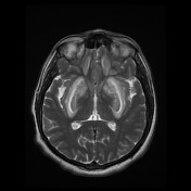
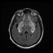

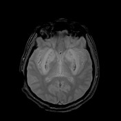
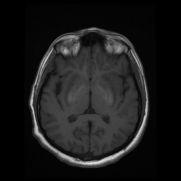
Bilateral basal ganglia symmetrical mixed-signal abnormalities with diffusion restriction and signal drop-in gradient-echo images mainly involving the putamen and head of the caudate nucleus.
Large bilateral frontal subcortical white matter signal abnormalities are also seen depicting diffusion restriction.
Small signal abnormalities are also seen in the occipital subcortical white matter.
Findings are indicating acute methanol intoxication with hemorrhagic necrosis in the basal ganglia.
Cerebellum and brain stem appear preserved.
Normal ventricular system and sulci
Case Discussion
This case illustrates bilateral basal ganglia involvement demonstrating water restriction on diffusion-weighted images and hyperintense signal on T1 indicating hemorrhagic necrosis. This patient present through the ER with methanol intoxication. Typical appearances of acute phase are illustrated by the involvement of the putamen with hemorrhagic necrosis and subcortical white matter of the frontal and occipital regions.


 Unable to process the form. Check for errors and try again.