
Presentation
G3P2 at 18 weeks. Routine ultrasound.
Patient Data
Age: 40 years
Gender: Female
From the case:
Autosomal recessive polycystic kidney disease - antenatal
Download
Info
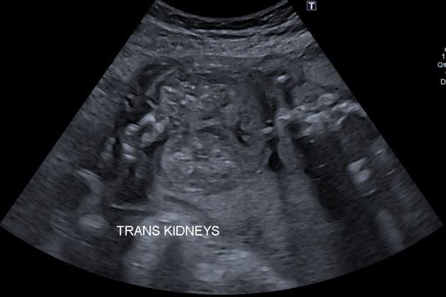
Enlarged kidneys, relatively echogenic, with loss of the corticomedullary differentiation. The reniform shape is preserved with associated oligohydramnios. The urinary bladder was not visualized which may indicate a lethal form of autosomal recessive polycystic kidney disease.
No other fetal anomalies were seen.
Case Discussion
Ultrasound faetures of an autosomal recessive polycystic kidney disease (ARPKD) with relatively severe oligohydramnios.


 Unable to process the form. Check for errors and try again.