
Presentation
Mechanical fall.
Patient Data

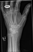
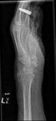
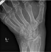
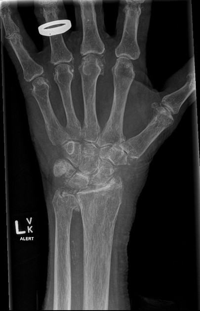
Severe osteopenia with cortical thinning. No malalignment or fracture. Calcific densities within the DRUJ, tri scaphe and radiocarpal joint spaces consistent with chondrocalcinosis. Hook osteophyte of the third metacarpal head. Vascular calcification.
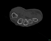

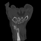
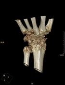
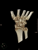
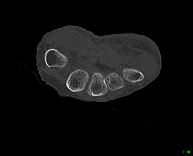
Extensive chondrocalcinosis throughout the wrist with soft tissue ossification and hook like osteophytes of the 2nd and 3rd metacarpals suggesting CPPD. Subchondral cysts and evident in the capitate, scaphoid, lunate, triquetrum and distal ulna.
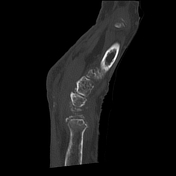
No fracture identified. Ossification adjacent to the radial styloid appears well corticated, unlikely to be related to an acute injury.
Wrist joint effusion present.
IMPRESSION
- No fracture identified.
- Extensive features of CPPD.
Case Discussion
Chondrocalcinosis and hook osteophytes are pathognomonic for CPPD.


 Unable to process the form. Check for errors and try again.