
Presentation
Abdominal pain.
Patient Data


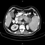


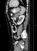
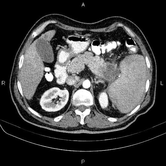
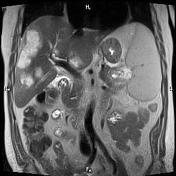
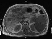
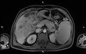
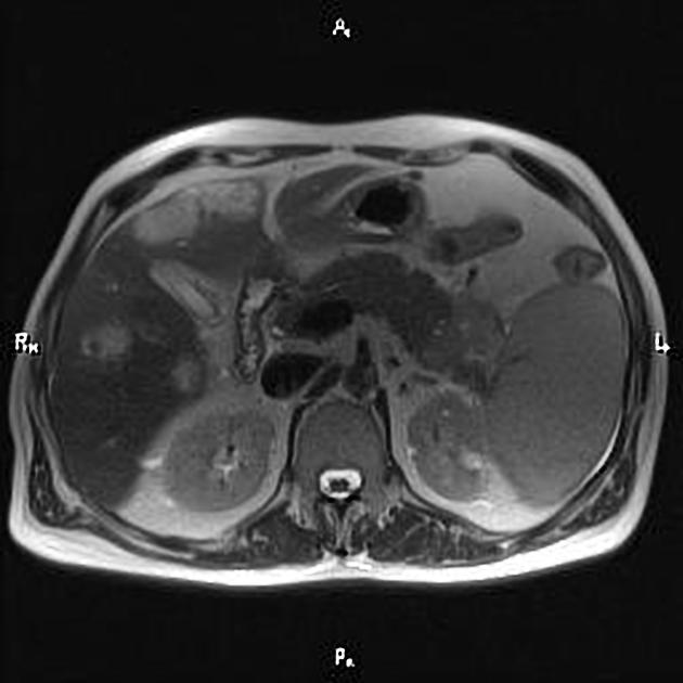
A 38mm low enhancing mass is seen at tail of pancreas. In addition, multiple low enhancing masses are seen in the liver parenchyma. The largest one is 37mm.
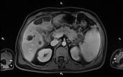
A few small cysts with smooth and thin walls, sharp and distinct marginations, and a homogenous water density were seen in left kidney. The largest one is 13mm in diameter.
Two tiny stone (≤4mm) were found in right renal calices.
The prostate gland is enlarged. Its volume measured about 43ml.

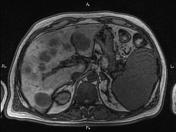
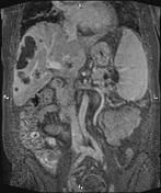
A 44×40mm heterogonous enhancing mass is noted at pancreas tail. Splenic vessels seems to be involved.
Multiple target shape masses are seen at liver, the largest one diameter is 54mm.
Gallbladder is contracted.
Mild splenomegaly is noted. A 16mm splenule is seen at lower aspect of spleen.
A few cortical cysts are seen at kidneys.
Body of left adrenal gland is dominant and shows heterogonous enhancement.
There is no lymphadenopathy at para-aorta.
The prostate gland is enlarged. Urinary bladder shows wall thickening.
There is no lymphadenopathy at pelvic cavity.
A few abnormal signal areas are seen at skeletal system.
Case Discussion
Metastatic pancreas mass; path proven pancreatic adenocarcinoma.


 Unable to process the form. Check for errors and try again.