
Presentation
Fall followed by anterior shoulder dislocation
Patient Data


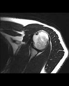
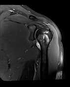

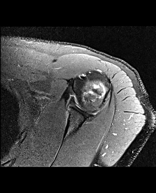
MRI study shows anterior glenolabral injury with torn anterior inferior labrum seen lifted from the edge of the glenoid with preserved attachment to the intact lifted periosteum from the anterior aspect of the glenoid. The labrum is not displaced
A localized cortical compression fracture is seen involving the posterolateral aspect of the humeral head with associated subcortical patchy bone marrow edema (Hill Sachs lesion)
Mild glenohumeral joint effusion distending the axillary recess
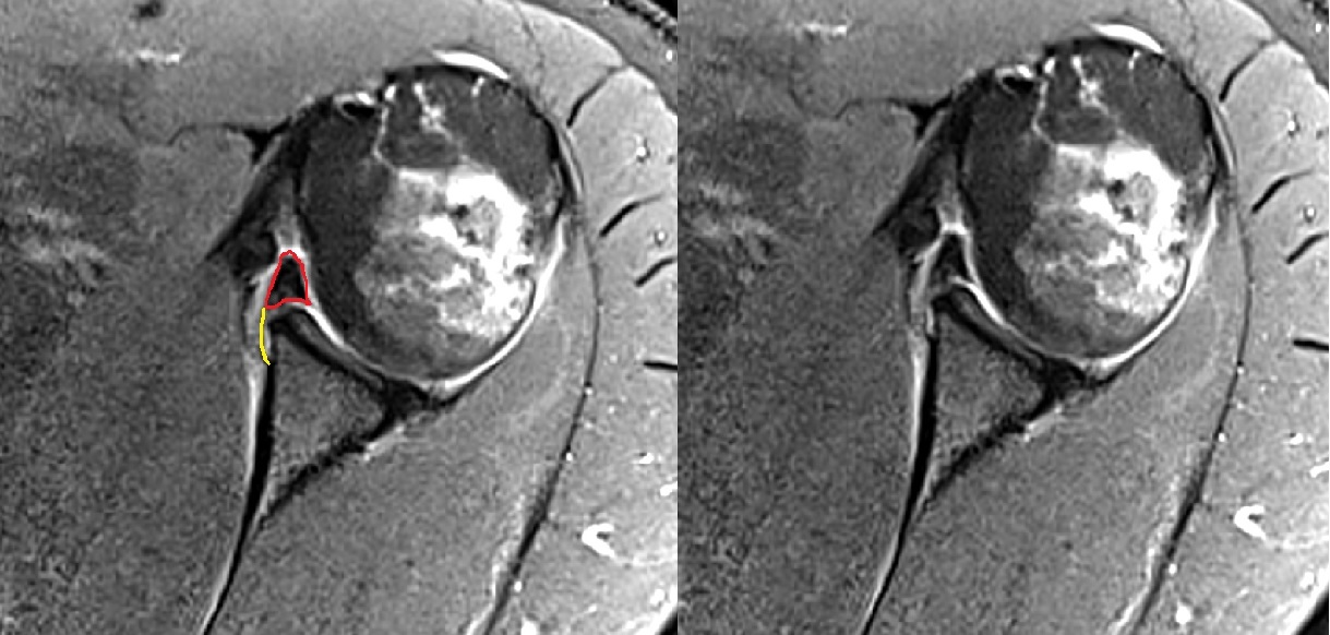
Annotated image highlighting the lesion. The red line outlines the anterior labrum and the yellow line outlines the intact lifted periosteum
Case Discussion
Here is a case of Perthes lesion one of the types of anterior glenolabral injuries. Others include Bankart, ALPSA, and GLAD lesions


 Unable to process the form. Check for errors and try again.