
Presentation
History withheld.
Patient Data


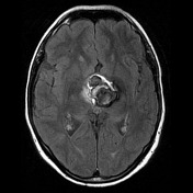
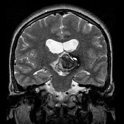

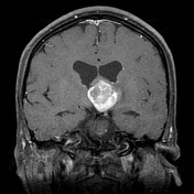
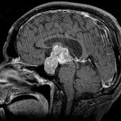
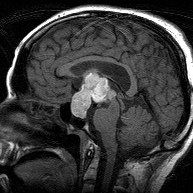
There is a large multi-lobulated mass with intrinsic high T1 signal and heterogeneous T2 signal, including large areas of signal drop out. The mass appears to arise from the pituitary fossa which is significantly expanded, and extend superiorly invaginating into the third ventricle. The outflow of the lateral ventricles is compromised with evidence of hydrocephalus and transependymal edema.
The optic chiasm is elevated and stretched over the anterior part of the mass, with the more posterior and superior component extending between the optic tracts.
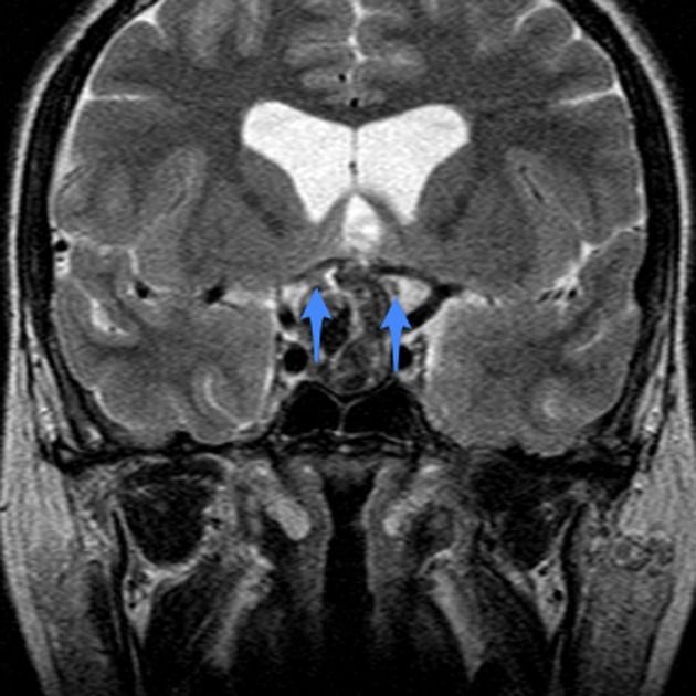
Optic nerves (green) lead back to the optic chiasm (red) which is markedly elevated and thinned. The superior part of the mass is located behind the chiasm, between the optic tracts (blue).
Case Discussion
This case dramatically demonstrates the appearances of pituitary apoplexy.


 Unable to process the form. Check for errors and try again.