
Presentation
Acute bilateral lower limbs weakness with foot drop.
Patient Data


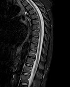
MRI study shows mild patchy signal changes of the distal spinal cord presents as intramedullary abnormal T2 hyperintensity with minimal swelling


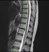
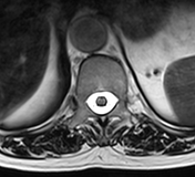
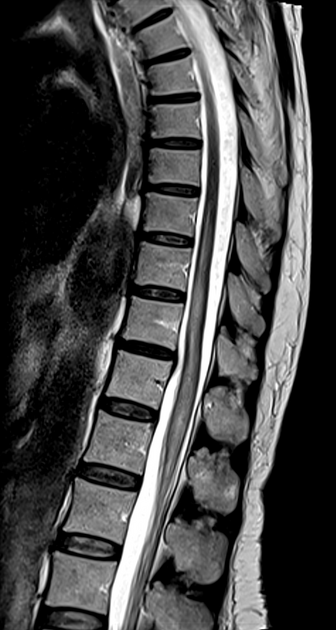
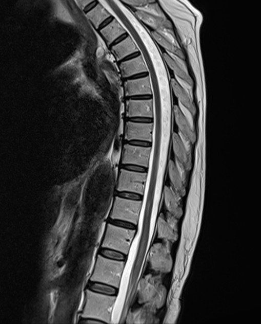
MRI study shows distal spinal cord long segment (4-5 vertebral levels) of signal alteration presents as a striking abnormal T2 hyperintensity with evident cord swelling. The disease is severe enough to involve most of the cross-sectional area of the cord
Case Discussion
Normal MRI doesn't exclude transverse myelitis as up to 40% of cases have no findings on MRI 1. In the early acute stage, the signal changes of the cord may be mild and questionable however, in the subacute stage, the cord changes appear evidently.
In this case, the neurologist's diagnosis according to the patient's history and examination was severe transverse myelitis secondary to acute viral infection, that was not correlated with the MRI except lately. Radiologists should be aware of this to avoid exclusion or underestimation of the cord disease, they should correlate with the clinical data as well


 Unable to process the form. Check for errors and try again.