
Presentation
Worsening disorientation. Multiple recent head traumas in history, prior CT scans showed no acute IC pathology. On long term anticoagulant therapy.
Patient Data


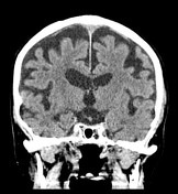
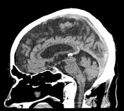
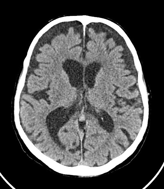
Advanced fronto-parietal dominant brain atrophy and chronic small vessel ischemic changes. Note lateral ventricle asymmetry. Thin, crescentic bilateral frontal subdural collections isodense to liquor. The cortical vein sign proves to be invaluable in outlining the boundary between the dilated subarachnoid space and the low density collections, and enables confident diganosis of bilateral subdural hygroma without iv. contrast in a patient with poor renal function.

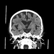
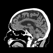
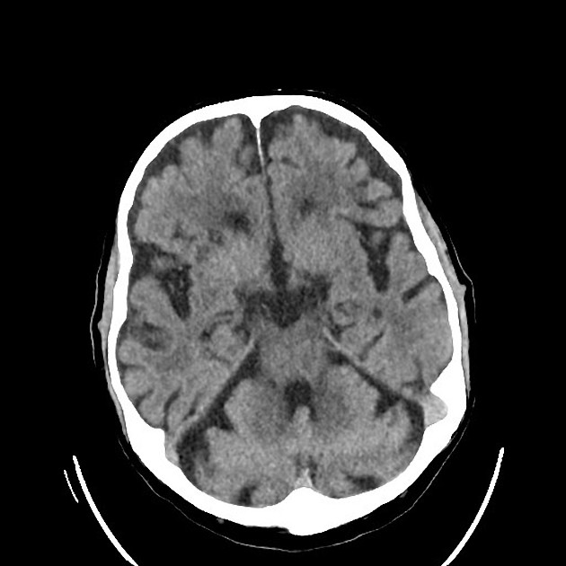
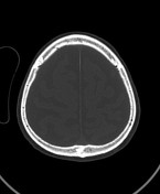
Striking difference when compared to available prior CT: cortical veins run adjacent to the inner table of the skull in the absence of hygroma.
Case Discussion
The case demonstrates the value of the cortical vein sign, and similarly to many other pathologies comparisons to recent prior imaging, in the diagnosis of subdural hygroma.


 Unable to process the form. Check for errors and try again.