
Cerebral autosomal dominant arteriopathy with subcortical infarcts and leukoencephalopathy (CADASIL)
Presentation
Generalized lethargy,minor visual disturbance recently;TIA ;collapse + seizure ? intracranial pathology.
Patient Data

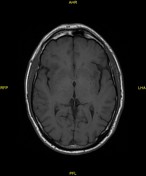
Extensive low attenuation in the periventricular white matter of both cerebral hemispheres with large confluent areas in the frontal lobes.
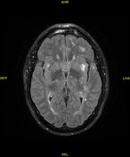

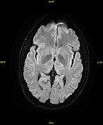
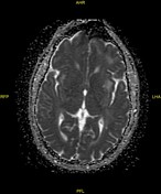
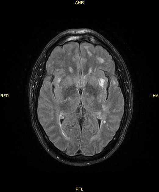
Extensive and in place in near confluent high T2 foci in the periventricular and deep white matter of both cerebral hemispheres, basal ganglia and internal/external capsules. No posterior fossa or callosal foci.
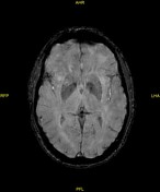
No diffusion restriction. No hemosiderin deposition.
Case Discussion
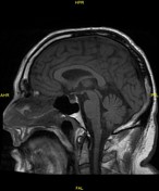
Widespread mature ischemic changes in the anterior circulation territories. Given the substantial temporal lobe and frontal lobe changes and the relatively young age of the patient, CADASIL is a strong possibility.
Ultrasound of the carotids was normal, with no evidence of atheromatous plaques.
The patient also exhibited many of the clinical features of CADASIL, such as features suggestive of multiple TIAs, a seizure and no history of atherosclerotic disease.


 Unable to process the form. Check for errors and try again.