
Presentation
Headaches.
Patient Data


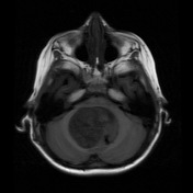
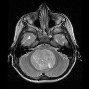
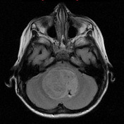

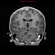
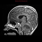
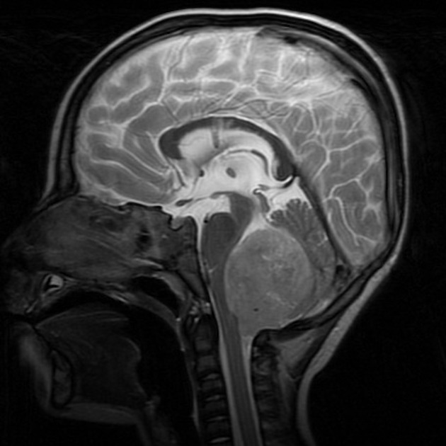
A very large rounded heterogenous tumor is present in the fourth ventricle, bulging inferiorly through the foramina of Luscka and Magendie. It is hyperintense on T2 weighted images, low on T1 weighted images, with heterogeneous contrast enhancement and has intermediate restricted diffusion on ADC. A ventricular shunt has been inserted through the right frontal lobe.
Case Discussion
This case illustrates the difficulty in categorically distinguishing medulloblastomas from ependymomas.
This child went on to have a craniotomy confirming the diagnosis of a posterior fossa ependymoma.
Note: as molecular profiling is unavailable in this case, it would have the diagnosis of posterior fossa ependymoma not otherwise specified (NOS).


 Unable to process the form. Check for errors and try again.