
Presentation
Poor urinary stream
Patient Data
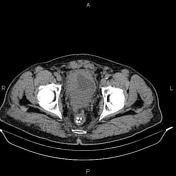

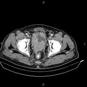


The prostate gland is enlarged and shows heterogeneous enhancement with irregular margin; infiltrated seminal vesicles, adjacent urinary bladder wall and distal of both ureters. There are multiple enlarged lymph nodes in the vicinity of the mass as well as bilateral para-iliac regions with maximum SAD of 23 mm.
Several mildly enlarged lymph nodes in the para-aortic regions.
Degenerative changes as osteophytosis are seen at the thoracolumbar spine.
Bilateral moderate hydroureteronephrosis. No renal or ureteral stones. A 7 mm cyst is present at segment 8 of the liver.
Case Discussion
Pathology proven case of prostate cancer with local invasion to seminal vesicles, urinary bladder and UVJs. Regional, parailiac and para-aortic lymphadenopathies.


 Unable to process the form. Check for errors and try again.