
Presentation
Heaviness in the pelvic region.
Patient Data
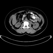

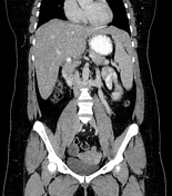
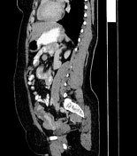

On the non-contrast study, there is a dilated tubular structure extending from the region of the left adnexa and terminating at the presumed site of the left renal vein, another tubular structure seen starting at the level of the left adrenal gland and again terminating at the level of the left renal vein.
On the post-contrast study, there is a retro-aortic left renal vein with subsequent dilatation of its proximal portion.
The tubular structure that extends from the pelvis is a thrombosed recanalized left ovarian vein. The vessel that extends from the left adrenal gland is the left adrenal vein.
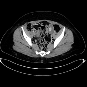
Note the pelvic varicosities.
Case Discussion
All findings seen in this case are in keeping with posterior nutcracker syndrome where the left renal vein is trapped by the aorta anteriorly and the spine posteriorly with resultant dilatation of its proximal portion and dilatation of all venous territories that drain into the left renal vein, namely the left gonadal (ovarian/testicular) vein and left adrenal vein.
Due to stagnation of flow inside these vessels, they are more liable to thrombosis with subsequent pelvic heaviness and varicosities. Recanalization is possible as seen in the left ovarian vein of this patient.
Case courtesy of Dr Alaa' Suoud, radiology specialist.


 Unable to process the form. Check for errors and try again.