
Presentation
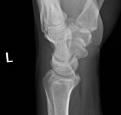
Post fall, ? injury of left wrist
Patient Data
Age: 45 years
Gender: Male
From the case:
Os hypolunatum

Download
Info
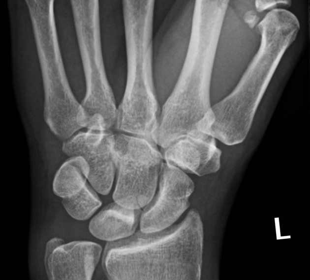
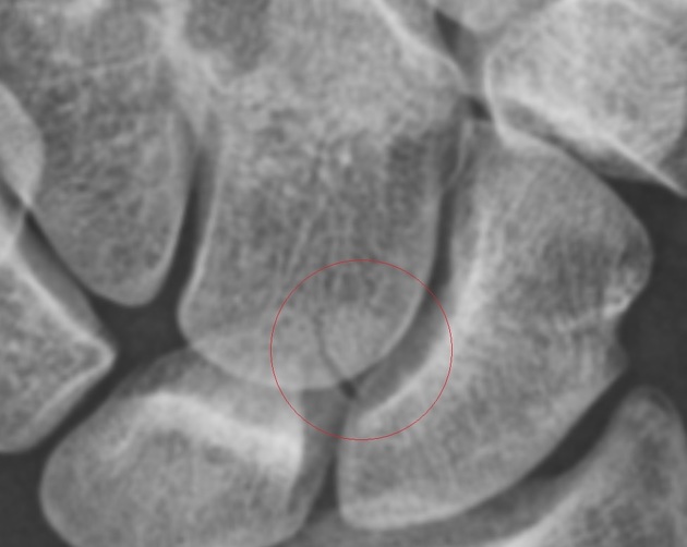
Small (5 x 5 mm) well-circumscribed normally-corticated bone at the lateral aspect of the distal lunate has the typical appearance of an os hypolunatum. No acute bony injury.


 Unable to process the form. Check for errors and try again.