
Presentation
Abdominal pain and palpable mass on physical exam.
Patient Data
Age: 55 years
Gender: Female
From the case:
Uterine leiomyosarcoma


Download
Info

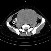
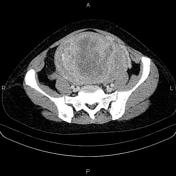
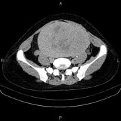
A 28 mm enhancing mass is noted at left side of para-aortic regions.
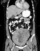
The uterus is enlarged. A 160×110 mm hetero enhancing mass is noted at uterine fundus.
There is no sign of extra uterus extension or local invasion to adjacent structures.
From the case:
Uterine leiomyosarcoma
Download
Info
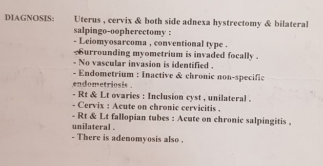
The patient underwent surgical resection and histopathology evaluation confirms leiomyosarcoma.
Case Discussion
Uterine leiomyosarcomas are malignant uterine tumors that arise from the myometrium. The uterus is the commonest location for a leiomyosarcoma.


 Unable to process the form. Check for errors and try again.