
Presentation
The patient was admitted to the ER, reporting a fall from the bike on the same day, complaining of pain in his elbow and left wrist.
Patient Data


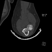
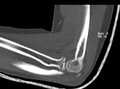
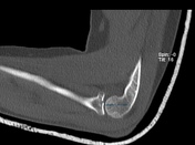

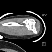
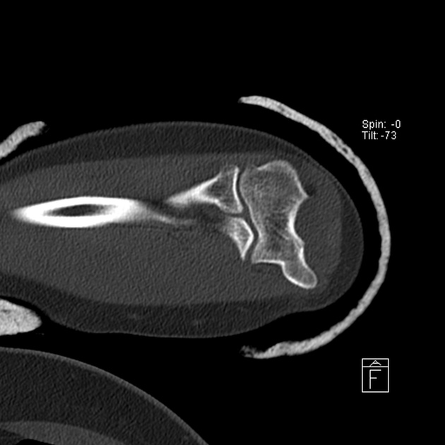
A vertical fracture line extending through the radial head's articular surface, with less than a 2 mm gap.
No other fracture was detected. The alignment of the elbow is intact.
Impression: Non-displaced intra-articular fracture of the radial head, consistent with a Mason type I fracture.
Case Discussion
Radial head fractures are a common type of elbow injury in adults 1-4. They usually occur during a fall on an outstretched arm with the forearm pronated and discrete flexion of the elbow joint 1-4. CT scan helps identify the location and size, number, morphology, articulate margin, fracture gap, and associated bone fractures and distinguishes the type of fracture in Mason classification 1-4. Radiologists should adopt the same terminology and classification systems used by orthopedic surgeons to enhance communication 1.
This case is a typical example of a non-displaced fracture of the radial head - Mason type 1, in which, in general, the treatment is conservative.


 Unable to process the form. Check for errors and try again.