
Presentation
Scrotal swelling of 6 years duration.
Patient Data

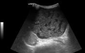

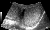
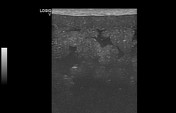
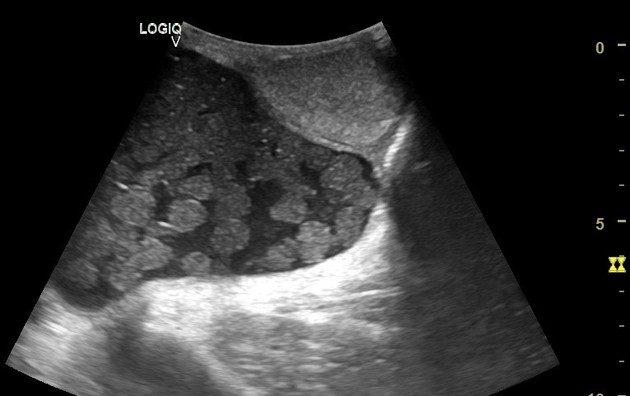
Scrotal ultrasound revealed 5.4 cm x 9.2 cm left side cystic lesion with multiple non dependent floating hyperechoic balls with marked posterior acoustic attenuation and foci of linear echogenicity surrounded by hypoechoic collection with low level internal echoes.
The lesion pushed the ipsilateral testis caudally.
Both testes are seen separately and have normal parenchymal echogenicity.
Color Doppler interrogation showed no intrinsic vascularity.
Case Discussion
Fine needle aspiration was carried out and cytology analysis showed keratinous thick fluid with squamous cells and fat suggesting the diagnosis of dermoid cyst and the patient has been referred for surgical management.
Inguinal dermoid cyst or mature cystic teratoma is a rare entity with few published case reports on English literature. These lesions are believed to arise from spermatic cord or round ligament. Floating intracystic echogenic fat balls, although not seen frequently, are considered characteristic for dermoid cyst.


 Unable to process the form. Check for errors and try again.