
Presentation
Two years history of unintentional movements with progressive rigidity. Family history of the same disease.
Patient Data
Age: 35 years
Gender: Female
From the case:
Huntington disease


Download
Info

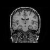
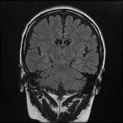
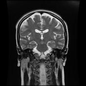
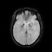
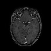
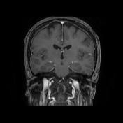
The MRI demonstrates:
- enlargement of the frontal horns of the lateral ventricles
- atrophy of the caudate nuclei
- prominent putaminal volume loss bilaterally
- blooming on eSWAN of the basal ganglia most likely due to iron deposition
From the case:
Huntington disease

Download
Info
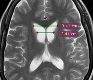
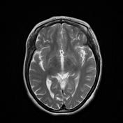
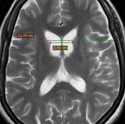
Annotated images:
- frontal horn width to intercaudate distance ratio (FH/CC) = 1.4 (normal range 2.2 to 2.6)
- intercaudate distance to inner table width ratio (CC/IT) = 0.21 (normal range 0.09 to 0.12)
Case Discussion
The clinical history and the MRI features are suggestive of Huntington disease.
Additional contributor: ZE. Boudiaf, MD, CHU Constantine, Algeria


 Unable to process the form. Check for errors and try again.