
Presentation
Trauma.
Patient Data
Age: 40 years
Gender: Male
From the case:
Isolated pisiform bone fracture (MRI)


Download
Info
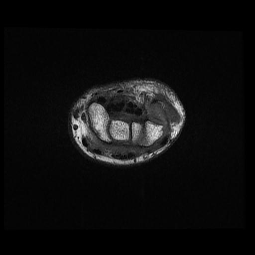
Fracture line and marrow edema signals within the pisiform bone
Bone marrow edema along with trapezium tubercle
Synovial cyst originated from triquetrum and pisiform bones joint space.
Case Discussion
A case of isolated pisiform bone fracture.


 Unable to process the form. Check for errors and try again.