
Presentation
Abdominal distention, constipation, and urge incontinence for two months. There is also history of uterine prolapse, generalized weakness and poor oral intake.
Patient Data

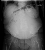
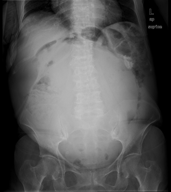
Sizable abdominopelvic soft tissue density lesion displacing the bowel loops to the periphery. Small focal calcified lesion in the left hypochondrium. No pneumoperitoneum is seen.
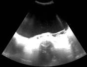
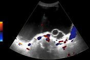
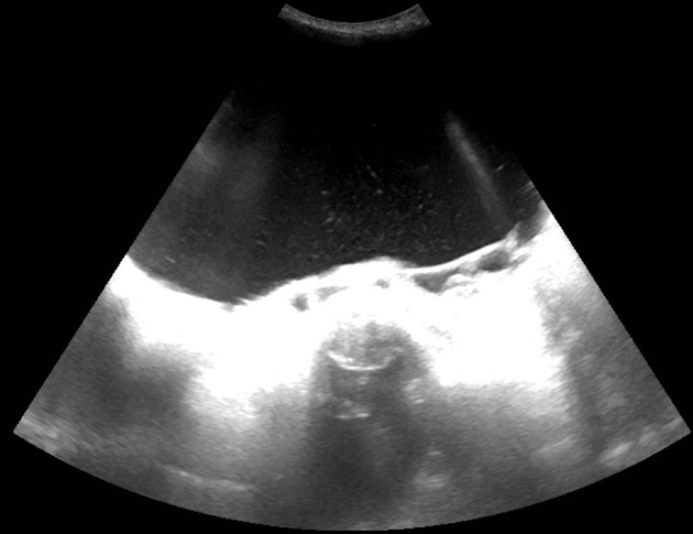
Huge abdominopelvic cystic lesion containing internal echoes and a few septations. No significant internal vascularity is seen in it.
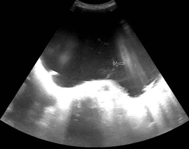
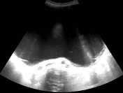
One of the thin separations is shown at the arrow.
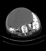

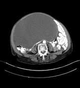
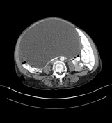
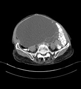
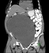
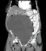
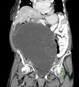
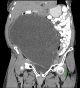
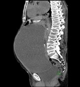
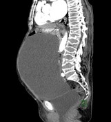

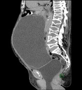

Huge abdominopelvic cystic mass (average density = 5 HU) measuring about 14 x 21 x 24 cm, likely arising from the right ovary. It has multiple thin peripheral enhancing septations; however,no enhancing solid component or calcifications are seen in it. A focal peritoneal calcific density lesion measuring 2.5 x 1.8 cm is seen in the left hypochondrium. Minimal free fluid identified in the right lumbar region. No significant lymphadenopathy or abdominal visceral metastases are seen. Prolapsed uterus and lower part of the urinary bladder associated with chronic bilateral moderate hydroureteronephrosis. Pooling of contrast noted in the renal calyceal system on delayed excretory phase. Diffuse osteopenia and multiple fish vertebra in the dorsolumbar spine.
Case Discussion
- Tumor markers: CA 19.9= <2 (≤37U/ml), CA 125=24 (≤35U/ml), CA 15.3=16.7 (≤31.3U/ml), CEA=9.3 (≤5ng/ml), LDH=249 (125-220 U/L).
- Procedure: Staging laparotomy, bilateral salpingo-oophorectomy (BSO), subtotal abdominal hysterectomy, omentectomy, lymph node sampling/biopsy and appendectomy.
Diagnosis:
- Right ovarian mass: Mucinous cystadenoma. Negative for malignancy. (Gross description: Huge gray-white cyst weighing 5200 grams and measuring 27 x 22 x 13.5 cm. The surface is grayish-white and glistening with numerous vessels showing and traversing the surface on the inner aspect. There is attached fallopian tube 8 cm long and 0.6 cm in diameter. Cyst opening released about 3000 ml of slightly turbid, but mostly clear fluid. The inner surface of the cyst shows reticulation and the cyst appears to be multilocular. No obvious gross solid area seen).
- Appendix: No significant abnormality.
- Lymph node sampling: Four reactive lymph nodes histologically identified. Negative for malignancy.
- 3 cm left hypochondrial calcified omental nodule: Dystrophic calcification. Negative for malignancy.
- Uterus with left tube and left ovary: Uterus shows one intramural leiomyoma. No significant abnormality seen in endometrium. Cervix showed active chronic cervicitis with no evidence of significant dysplasia. Left ovary and fallopian tube show only paratubal cysts.


 Unable to process the form. Check for errors and try again.