
Presentation
Headaches and visual disturbance.
Patient Data


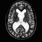
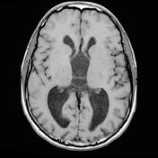


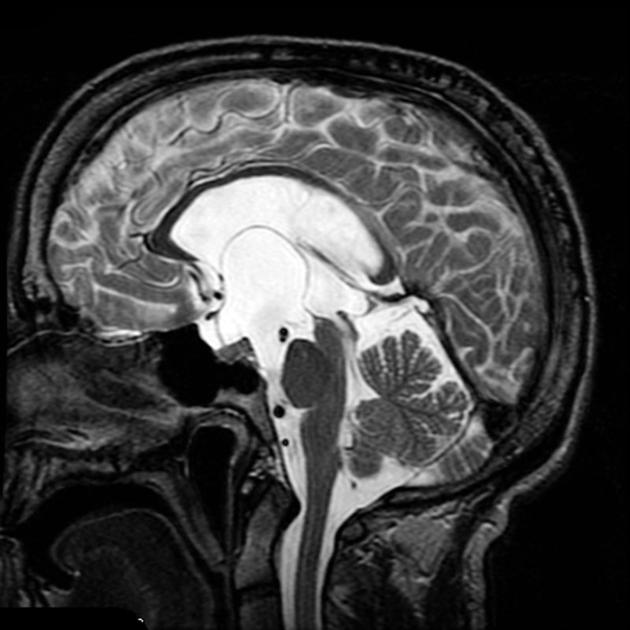
MRI demonstrates a CSF intensity space distorting the optic chiasm and pituitary infundibulum (pushing them forwards and upwards). A thin membrane can be seen (best on sagittal and axial T2 weighted images) invaginating upwards into the third ventricle, splaying the septum pellucidum and even bulging into the left foramen of Munro. The lateral ventricles are dilated, in keeping with obstructive hydrocephalus, clearly long standing (note large size and lack of transependymal edema).
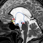
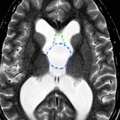
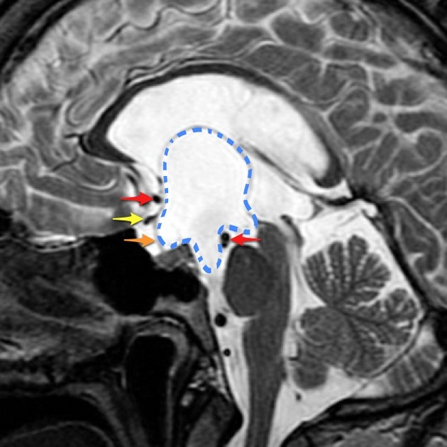
Anterior cerebral arteries and basilar artery tip are seen as flow voids (red arrows); optic chiasm (yellow arrow); pituitary infundibulum (orange arrow); septum pellucidum (green dotted lines); arachnoid cyst (blue dotted line).
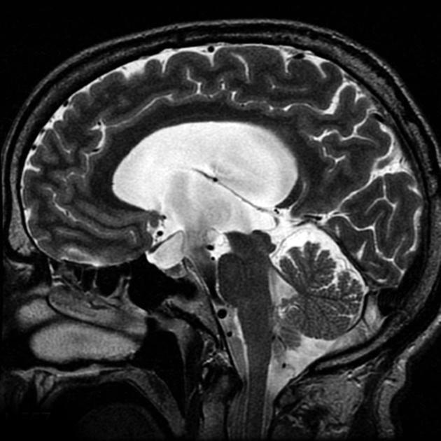
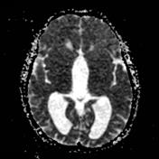
A third ventriculostomy was performed, and the floor of the cyst was also opened into the interpeduncular cistern. Prominent CSF pulsation can be seen continuously from the third ventricle to the prepontine cistern.
Note the reduced distortion on the optic chiasm and pituitary infundibulum.
Case Discussion
Suprasellar arachnoid cysts have historically been difficult to diagnose, although with modern MRI the diagnosis is more straight forward.


 Unable to process the form. Check for errors and try again.