
Presentation
Abdominal pain.
Patient Data
Age: 55 years
Gender: Female
From the case:
Pancreatic adenocarcinoma

Download
Info

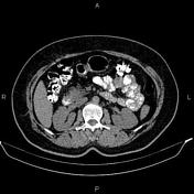
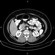
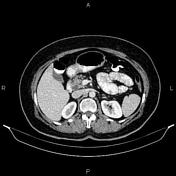
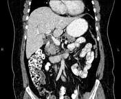
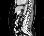
Mild dilatation of intrahepatic and extrahepatic bile ducts was seen. CBD measured 12mm in caliber.
A 21 mm iso dense mass was seen in uncinate process of pancreas, which enhances less than normal pancreatic parenchyma on post-contrast images.
Case Discussion
Pathology proven case of pancreatic adenocarcinoma with mild cholestasis.
No local invasion, No regional lymphadenopathy, No detectable metastasis.


 Unable to process the form. Check for errors and try again.