
Presentation
Right-sided neck swelling.
Patient Data
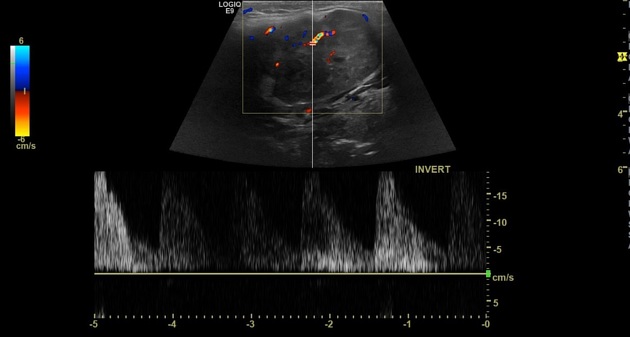
Ultrasound study shows right submandibular well-defined ovoid lobulated soft tissue mass lesion measures about 55 x 37 x 31 mm, it presents heterogeneous hypoechogenicity with small areas of cystic changes and posterior acoustic enhancement, no calcifications, it shows internal vascularity on the color Doppler study.
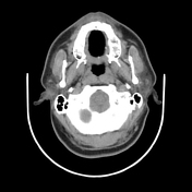

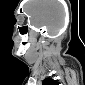
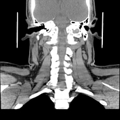

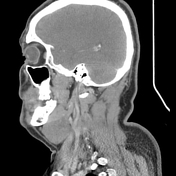
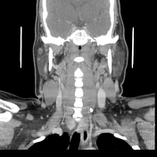

CT study shows right submandibular soft tissue mass lesion inseparable from the right submandibular gland, abutting the anterior aspect of right sternomastoid muscle with clear surrounding fat planes. It elicits slight hypodensity on non-enhanced CT study and mild heterogeneous enhancement on the postcontrast study with few foci of cystic breakdown. No calcifications are seen.
Bilateral submandibular and deep cervical multiple small lymph nodes.

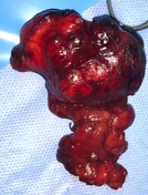
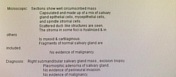
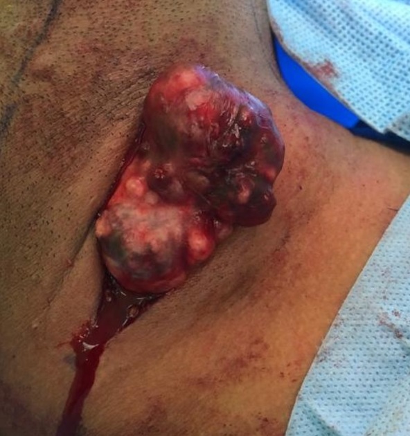
Surgical excision of the mass and the right submandibular gland was done. Histopathology revealed pleomorphic adenoma with no evidence of malignancy.
Case Discussion
Pleomorphic adenomas of the salivary glands, also known as benign mixed tumors, are the most common salivary gland tumors.
On CT, when small, they have homogeneous attenuation and prominent enhancement. When larger, they can be heterogeneous with less prominent enhancement, foci of necrosis, and possible delayed enhancement. Small regions of calcification are common
This case is an example of a large pleomorphic adenoma
Additional contributor from the radiologist Dr Somia Elbadawy
Intraoperative photos are courtesy of Dr Adel Abdelwahed, the oncology surgery consultant


 Unable to process the form. Check for errors and try again.