
From the case:
Left sided superior vena cava
Download
Info
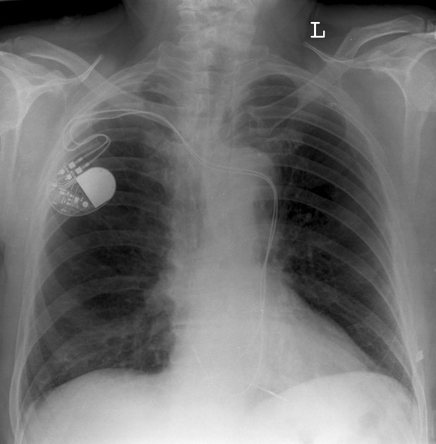
Frontal X-ray of the chest demonstrates the two wires from the right-sided permanent pacemaker wires passing to the left of the midline before descending posterior to the heart. This is consistent with a left-sided SVC.
Case Discussion
Left sided SVC is one of the more frequent caval abnormalities


 Unable to process the form. Check for errors and try again.