
Presentation
Sudden severe lower abdominal pain associated with vomiting 1 day. No change in the urinary or bowel habits.
Patient Data

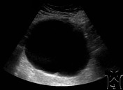
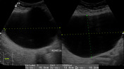


Large well-defined anechoic thin walled cyst with clear fluid contents, measuring 15 x 13 x 10 cm seen in the infra-umbilical region. No septations, calcifications, solid component or internal vascularity is seen in it.
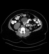

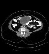
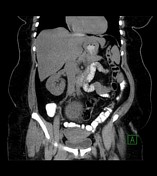
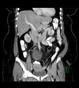
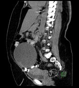
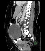
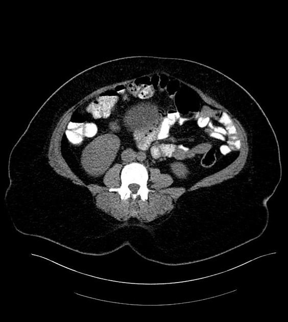
Large, well-defined, cystic lesion containing clear/simple fluid contents, arising from the pelvis and extending upwards into the lower abdomen, measuring 16 x 14 x 11 cm. A shallow indentation is seen at its left inferior surface. The cyst wall is relatively thin and regular (less than 4 mm thickness), and has no solid mural nodules or papillary projections. The inferior root of the lesion is inseparable from the right ovary, fallopian tube and broad ligament. Surrounding fat planes are clear. A few small radiopaque gallstones. Two lobulated thin-walled pancreatic cysts, measuring 2 x 2 cm and 1.3 x 1.1 cm, are seen. No ascites or lymphadenopathy is seen.
Case Discussion
Procedure: Laparotomy with right salpingo-oophorectomy.
Diagnosis: Benign serous cystadenoma.
Gross Description: Specimen submitted in one formalin container labeled “Right ovarian cyst”. It is composed of a thin-walled oval cyst measuring 17 x 14 x 10 cm and weighing 1353 grams. On opening up, it is filled with clear, yellowish watery fluid. The inner lining is smooth without any papillation.


 Unable to process the form. Check for errors and try again.