
From the case:
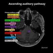
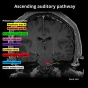
Ascending auditory pathway (annotated MRI)


Download
Info

Annotated ascending auditory pathway anatomy on T1 and T2W MRI of a patient with normal imaging findings.
Case Discussion
Ascending auditory pathway (in order from 1 to 7) 1-3:
Cochlear nerve at internal auditory canal
Cochlear nucleus at pontomedullary junction/upper medulla
Superior olivary complex at lower pons
Lateral lemniscus at pons (named white matter tract)
Inferior colliculus at tectum
Medial geniculate body at thalamus
Primary auditory cortex (Brodmann areas 41 and 42) at superior temporal gyrus


 Unable to process the form. Check for errors and try again.