
From the case:
Cerebellar vermis (illustration)

Download
Info

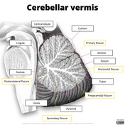
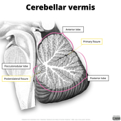
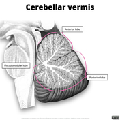
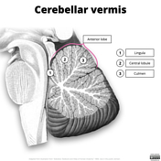
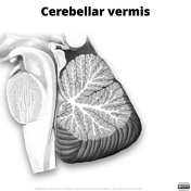
Illustration depicting the parts of the cerebellar vermis in the sagittal plane. It should be noted that there appears to be high variability in how this is depicted. The labels have been adapted from a combination of Sobotta's original labels and the 37th edition of Gray's Anatomy.
Adapted from an illustration from “Sobotta's Textbook and Atlas of Human Anatomy” 1908, now in the public domain.
Original image obtained from Wikimedia Commons here.
Touch-ups and labels by Frank Gaillard


 Unable to process the form. Check for errors and try again.