Presentation
Gradual abdominal distension.
Patient Data
![](https://prod-images-static.radiopaedia.org/images/55271959/013_thumb.jpeg)
![This study is a stack](/packs/stack-YQKLCKBI.gif)
![](https://prod-images-static.radiopaedia.org/images/55271983/013_thumb.jpeg)
![This study is a stack](/packs/stack-YQKLCKBI.gif)
![](https://prod-images-static.radiopaedia.org/images/55272031/013_thumb.jpeg)
![This study is a stack](/packs/stack-YQKLCKBI.gif)
![](https://prod-images-static.radiopaedia.org/images/55272189/011_thumb.jpeg)
![This study is a stack](/packs/stack-YQKLCKBI.gif)
![](https://prod-images-static.radiopaedia.org/images/55272211/013_thumb.jpeg)
![This study is a stack](/packs/stack-YQKLCKBI.gif)
![](https://prod-images-static.radiopaedia.org/images/55272235/013_thumb.jpeg)
![This study is a stack](/packs/stack-YQKLCKBI.gif)
![](https://prod-images-static.radiopaedia.org/images/55272259/013_thumb.jpeg)
![This study is a stack](/packs/stack-YQKLCKBI.gif)
![](https://prod-images-static.radiopaedia.org/images/55272298/010_thumb.jpeg)
![This study is a stack](/packs/stack-YQKLCKBI.gif)
![](https://prod-images-static.radiopaedia.org/images/55272355/049_thumb.jpeg)
![This study is a stack](/packs/stack-YQKLCKBI.gif)
![](https://prod-images-static.radiopaedia.org/images/55272459/013_thumb.jpeg)
![This study is a stack](/packs/stack-YQKLCKBI.gif)
![](https://prod-images-static.radiopaedia.org/images/55272480/010_thumb.jpeg)
![This study is a stack](/packs/stack-YQKLCKBI.gif)
![](https://prod-images-static.radiopaedia.org/images/55272499/011_thumb.jpeg)
![This study is a stack](/packs/stack-YQKLCKBI.gif)
![](https://prod-images-static.radiopaedia.org/images/55271959/013_big_gallery.jpeg)
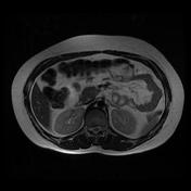
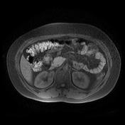
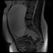
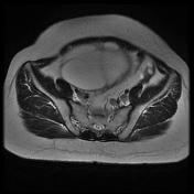
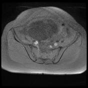
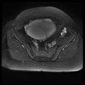
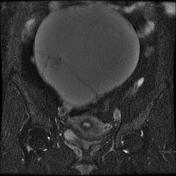
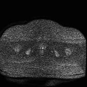
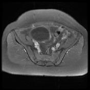
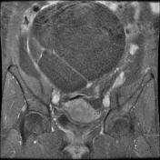
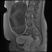
There is a 90*170*180 mm large midline multiseptate cystic lesion that elicits low on T1 and bright T2 fat sequence without water restriction that extends superiorly above the level of the umbilicus
It shows internal enhancing septations (measuring about 3 mm thickness) with no evidence of papillary projection or soft tissue components
The right ovary couldn’t be defined separately than mentioned cystic lesion
There are no signs of local invasion to adjacent structures and no regional lymphadenopathies
The left ovary is unremarkable
No free fluid is seen in cul–de–sac space.
Case Discussion
Pathology proved the case of ovarian serous cystadenoma which is a benign type of ovarian epithelial tumor at the benign end of the spectrum of ovarian serous tumors.