
Presentation
Right-sided hip pain.
Patient Data
Age: 60 years
Gender: Male
From the case:
Incidental prostate cancer on hip MRI


Download
Info
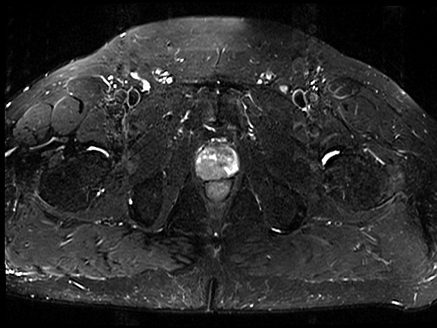
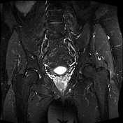
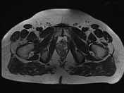
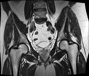
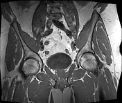
Moderate degenerative changes of both hips and the depicted lumbar spine (not all sequences shown).
Hypointense, "erased charcoal-like" (though the term is likely not appropriate to be used in a hip MRI) area measuring about 25 mm, mainly in the right peripheral zone of the prostate. The lesion is best depicted on STIR.
Case Discussion
The possibility of malignancy was raised, and the biopsy subsequently confirmed prostate cancer.
Musculoskeletal MRI exams in the region of the pelvis are high yield in terms of important incidental findings. Care must be taken to scrutinize the depicted urogenital organs, and the visualized segments of the GI tract.


 Unable to process the form. Check for errors and try again.