
Presentation
Work up for abdominal pain.
Patient Data
Age: 30 years
Gender: Male
From the case:
Duplicated inferior vena cava

Download
Info
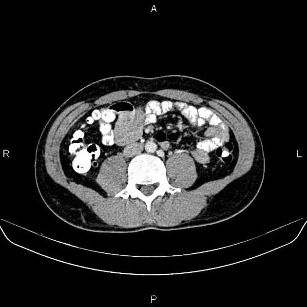
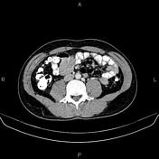
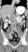
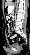
There is duplicated left sided inferior vena cava which crosses anterior to the aorta at the level of the left renal vein to join right IVC.
Case Discussion
Duplication of the inferior vena cava is a relatively rare vascular anomaly, but this caval abnormality needs to be recognized, especially in association with renal anomalies like crossed fused ectopia or circumaortic renal collar.


 Unable to process the form. Check for errors and try again.