
Presentation
Painful swelling of the left iliac region.
Patient Data


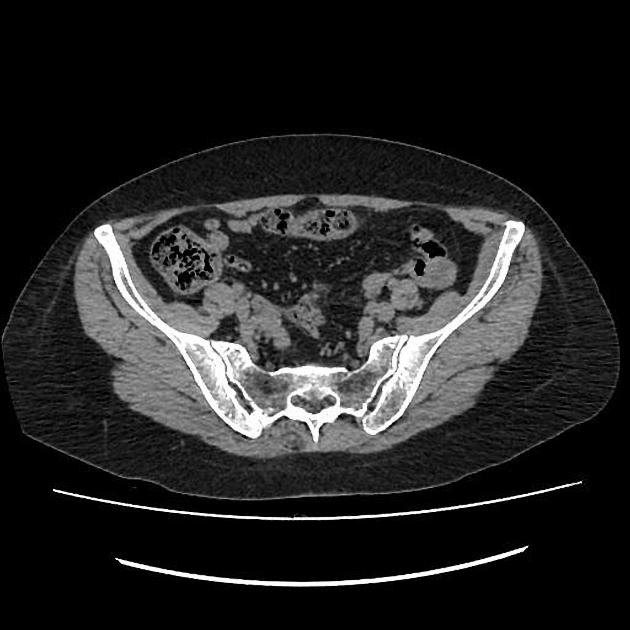
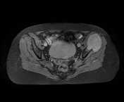
There is an osteolytic bone lesion centered on the left iliac wing with a soft tissue component measuring 55 x 50 x 45 mm, isodense to the muscles with vivid heterogeneous enhancement on postcontrast images.
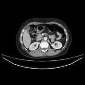
The rest of the chest and abdominopelvic CT is unremarkable except small simple right renal cyst. Lungs were clear (lung window not shown).
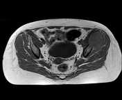
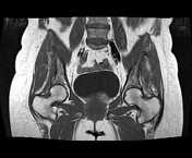
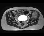
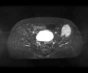
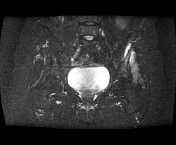
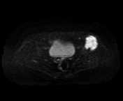
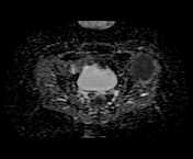
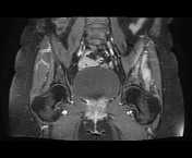
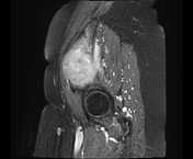
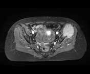

On MRI sequences the soft tissue component appears relatively isointense to the muscles on T1, high signal on T2 and STIR with heterogeneous enhancement following IV contrast and restricted diffusion on DWI/ADC. The iliacus and gluteus muscles are probably invaded. The hip joint is preserved.
Case Discussion
CT and MRI features of an osteolytic tumor centered on the left iliac wing with a soft tissue component invading probably the iliacus and gluteus muscles, pathologically proven as an Ewing sarcoma.


 Unable to process the form. Check for errors and try again.