
Presentation
Right hip pain
Patient Data
Age: 70 years
Gender: Female
From the case:
Osteoarthritis of the hip
Download
Info
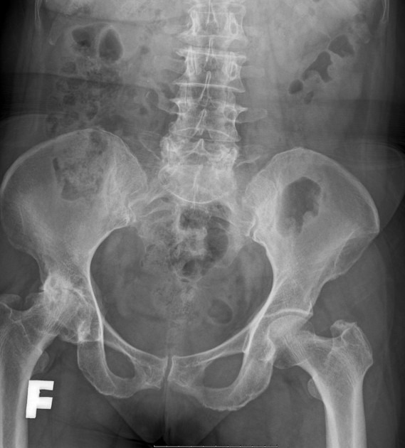
The right hip demonstrates asymmetric narrowing of the joint space, particularly at the weight-bearing segment, formation of marginal osteophytes, subchondral sclerosis, geodes, remodeling of the articular surface.
From the case:
Osteoarthritis of the hip


Download
Info

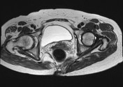
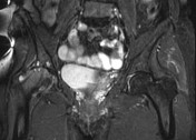
MRI findings are similar to X-rays. In addition, there is the eroded cartilage joint, joint effusion, and bone marrow edema in the right femoral head, acetabulum.
Case Discussion
X-ray and MRI findings are in keeping with osteoarthritis of the hip.


 Unable to process the form. Check for errors and try again.