
Presentation
Dyspnea on exertion. Coronary artery bypass grafts.
Patient Data
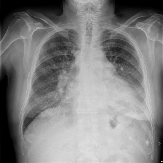
Cardiomegaly, peribronchial cuffing, ill-defined central/lower zone predominant opacity, subtle Kerley B lines and small left pleural effusion indicative of heart failure and pulmonary edema.
Additional border-forming structure lateral to the aortic arch.
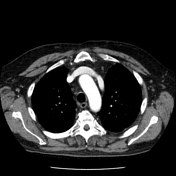

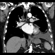
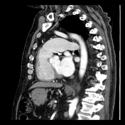

Aberrant left superior pulmonary vein draining into the left brachiocephalic vein.
A mildly dilated right ventricle and proximal pulmonary arteries.
LIMA to LAD and saphenous vein to PDA grafts.
Left pleural effusion and lower lobe collapse/consolidation.
Case Discussion
Echocardiogram: There is mild tricuspid incompetence into a mild-to-moderately dilated right atrium. The pulmonary systolic pressure is estimated at 37mmHg plus right atrial pressure.
Partial anomalous pulmonary venous return is a cause of left to right shunting and the increased flow can cause dilatation of the corresponding blood vessels and heart chambers as well as pulmonary hypertension. Symptoms include dyspnea.
In this case, the aberrant left superior pulmonary vein is border-forming on the chest radiograph.


 Unable to process the form. Check for errors and try again.