Presentation
Recurrent attacks of upper abdominal pain. Incidentally discovered hepatic focal lesion in ultrasound.
Patient Data
![](https://prod-images-static.radiopaedia.org/images/55669438/0._big_gallery.jpeg)
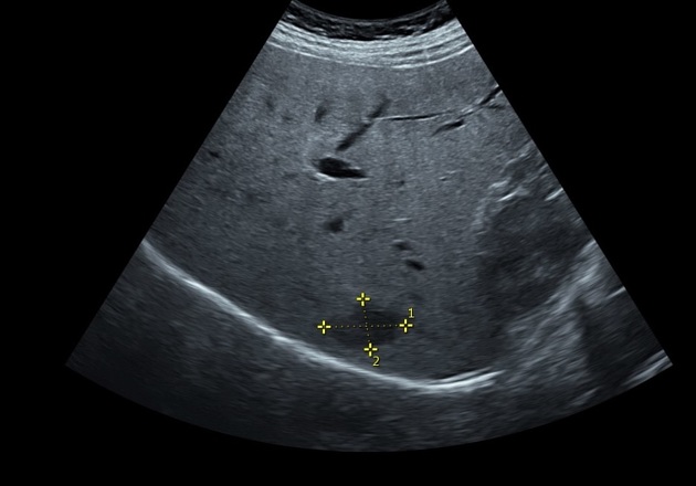
Ultrasound study shows bright fatty liver, small hypoechoic hepatic focal lesion at segment VII, gallbladder stones, and left ovarian small simple cyst.
![](https://prod-images-static.radiopaedia.org/images/55669471/4ddb415242e65d19acacba378e4387caa23bfdab797109f7c9e067e5081b799d_thumb.jpeg)
![This study is a stack](/packs/stack-YQKLCKBI.gif)
![](https://prod-images-static.radiopaedia.org/images/55669560/0a447684914d106554c44e7e92d2973a3c9f4b11b2772f2508e9c23575e7d69f_thumb.jpeg)
![This study is a stack](/packs/stack-YQKLCKBI.gif)
![](https://prod-images-static.radiopaedia.org/images/55669695/ec31da336e487788891eeed5aa91b39dd46d8839329b3bc2b145b0eec4de91ed_thumb.jpeg)
![This study is a stack](/packs/stack-YQKLCKBI.gif)
![](https://prod-images-static.radiopaedia.org/images/55669796/9c27d7ec4a08b3e96a5ebfe1fb16270711395109d9e2e061c8641728f7dec757_thumb.jpeg)
![This study is a stack](/packs/stack-YQKLCKBI.gif)
![](https://prod-images-static.radiopaedia.org/images/55669945/ef33f7f87220c9b4bdd68942012fb278393d294f83af938103b599201d6650f6_thumb.jpeg)
![This study is a stack](/packs/stack-YQKLCKBI.gif)
![](https://prod-images-static.radiopaedia.org/images/55669992/0a1cef7a557c178f96ddfc483326ea335c8ccdd12e0ef60026d2b88fee16da55_thumb.jpeg)
![This study is a stack](/packs/stack-YQKLCKBI.gif)
![](https://prod-images-static.radiopaedia.org/images/55669877/a1cd7ea3f576487ace4e27418b35127ca2547f500a48e53ac24a9103551b49f4_thumb.jpeg)
![This study is a stack](/packs/stack-YQKLCKBI.gif)
![](https://prod-images-static.radiopaedia.org/images/55669471/4ddb415242e65d19acacba378e4387caa23bfdab797109f7c9e067e5081b799d_big_gallery.jpeg)
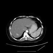
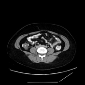
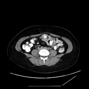
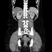
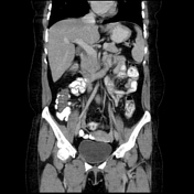
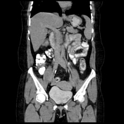
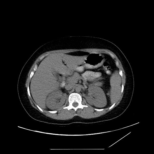
CT study shows fatty liver with segment VII small focal lesion measures 15x25 mm elicits isodensity in the non-contrast phase, early enhancement in the arterial phase that fades in the subsequent phases following the density of the blood pool (aorta). The lesion is isodense in the delayed phase with no washout.
Mildly thickened gallbladder wall and left adnexal cyst measures 2.5x4.5 cm are also noted.
![](https://prod-images-static.radiopaedia.org/images/55670022/0._thumb.jpeg)
![](https://prod-images-static.radiopaedia.org/images/55670023/0._thumb.jpeg)
![](https://prod-images-static.radiopaedia.org/images/55670024/0._thumb.jpeg)
![](https://prod-images-static.radiopaedia.org/images/55670025/0._thumb.jpeg)
![](https://prod-images-static.radiopaedia.org/images/55670022/0._big_gallery.jpeg)
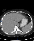
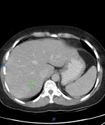
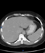
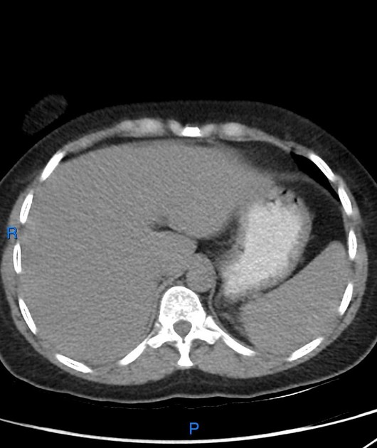
The annotated images highlight the lesion in different CT study phases.
Case Discussion
Flash filling hepatic hemangiomas are a type of atypical hepatic hemangioma that shows a quick homogeneous enhancement in the arterial phase and retains the contrast and remains isodense to the adjacent vascular pool in the rest of the phases. This is just a contrast fade and not a washout as the lesion never presents hypodensity relative to the hepatic parenchyma.
Hepatic hemangiomas are typically hyperechoic lesions on ultrasound. However, in a small proportion (10%) they are hypoechoic, which may be due to a background of hepatic steatosis, where the liver parenchyma itself is of increased echogenicity.
Ultrasound contribution by Dr. Shaimaa Hussain