
Presentation
Pelvic pain and palpable mass on physical exam.
Patient Data


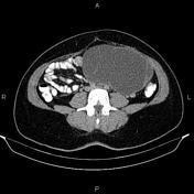
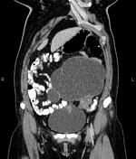
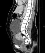
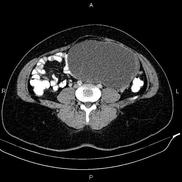
A 195 x 110 x 130 mm multiloculated cystic lesion without enhancing solid component is present at left adnexa, seeming to originate from the left ovary.
A small amount free fluid is observed at posterior cul-de-sac.
The uterus contains a few small fibroids.
The gallbladder is not seen at anatomical location due to prior resection.
Case Discussion
Left adnexal cystic lesion - pathology-proven ovarian serous cystadenoma which is a type of benign ovarian epithelial tumor at the benign end of the spectrum of ovarian serous tumors.
On CT, ovarian serous cystadenoma is often seen as a unilocular (typically) or multilocular cystic mass with homogeneous attenuation, with a thin regular wall or septum and usually no solid component or mural node.


 Unable to process the form. Check for errors and try again.