
From the case:
Submandibular duct stone
Download
Info
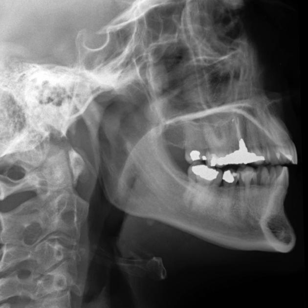
Plain film demonstrates an ovoid calcific density just below the angle of the mandible.
From the case:
Submandibular duct stone
Download
Info
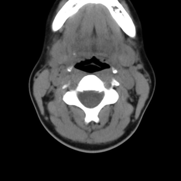
CT confirms the presence of calcific density on the left in a location likely to place it within the submandibular duct near the gland. The submandibular gland appears unremarkable (not enlarged; no surrounding stranding).
From the case:
Submandibular duct stone


Download
Info

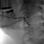
A sialogram confirms the presence of a submandibular duct stone located at the hilum of the gland.
Case Discussion
The duct was subsequently cannulated and the stone removed fluoroscopically.
Case courtesy of Bob Cook, MD. Western Memorial Regional Hospital Corner Brook, Newfoundland.


 Unable to process the form. Check for errors and try again.