
Presentation
Back pain, with increased inflammatory markers. Lumbar spine XR to assess for discitis.
Patient Data
Age: 50 years
Gender: Male
From the case:
Ruptured abdominal aortic aneurysm

Download
Info
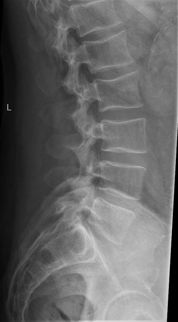
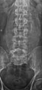
Normal included spine and pelvis.
Large peripherally calcified fusiform midline structure.
From the case:
Ruptured abdominal aortic aneurysm


Download
Info
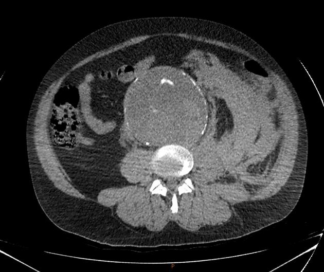
Infrarenal aortic aneurysm, maximally measuring 10.8 cm.
Left retroperitoneal stranding indicating aneurysm rupture. No active leak on arterial or delayed venous phase imaging (not included).
This also demonstrates the draped aorta sign - with the posterior wall of the aorta following the contour of the adjacent vertebral body as it decompresses into the soft tissues.
Case Discussion
This was an unexpected finding for the clinical team, and was urgently assessed by imaging and underwent open operative fixation that day.


 Unable to process the form. Check for errors and try again.