
Presentation
Postmenopausal bleeding
Patient Data
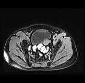

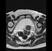
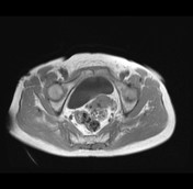
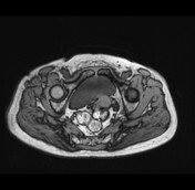
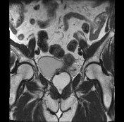
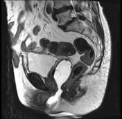
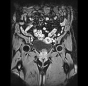
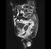
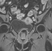

There is a mass in the endometrial cavity. It shows heterogeneous high signal on T2, heterogeneous low signal on T1 compared with myometrium, enhances less than myometrium on T1C+; a single feeding vessel extends to the mass consistent with pedicle artery sign.
The lesion appears to invade the anterior myometrial wall.
The uterus presents atrophy change.
The anterior abdominal wall hernia is seen.


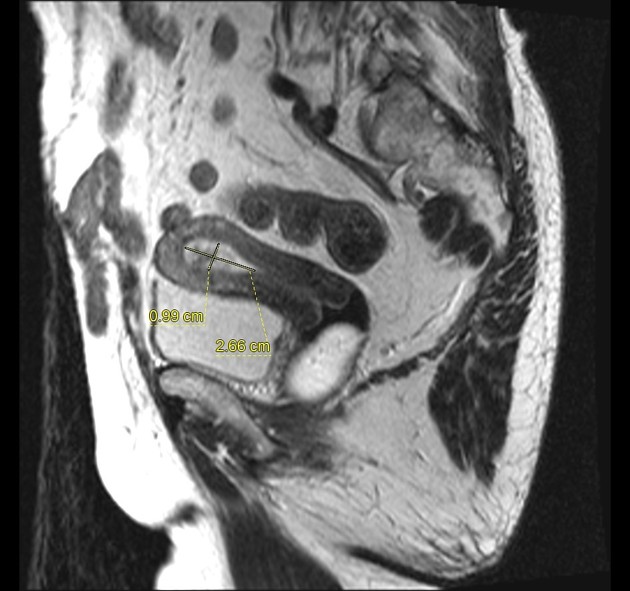
The mass measuring 10x26 mm
Yellow arrow: pedicle artery
The hysterectomy was performed, histopathology confirms the benign polyp of the endometrium.
Case Discussion
This case demonstrates the imaging features of endometrial polyps with pedicle artery sign on MRI.
The differential diagnosis includes endometrial hyperplasia, endometrial carcinoma.
The pedicle artery sign on transvaginal color Doppler sonography has a sensitivity of 86.67%, an accuracy of 86.67%, a positive predictive value of 100%, and no false positive. It improves the specificity for the diagnosis of endometrial polyps 1.


 Unable to process the form. Check for errors and try again.